
Musculoskeletal imaging in sports medicine is an evolving and exciting radiology sub-specialty. Driven as much by advanced technology as by improved perception of sporting injuries, it helps minimise injuries through timely diagnosis and correct treatment
Plain Radiographs and CT
Plain x-ray is often the initial imaging technique to investigate a sports related injury. Two orthogonal x-rays provide a comprehensive anatomical view economically and with lower radiation. Combined with clinical examination, plain x-rays help diagnose and treat sports related injuries.
Musculoskeletal trauma could also be assessed using CT for certain indications like complex fractures like subtle scaphoid and acetabular fractures. At Kokilaben Hospital, the studies are performed on a multidetector row dual source scanner with post processing to create multiplanner and 3D reconstructions that allow comprehensive assessment of the bony anatomy and pathology.
Ultrasound
Musculoskeletal ultrasound is a non-invasive, easily available, versatile and fairly accurate technique for the diagnosis of sports injuries. Some common indications include rotator cuff pathology, carpal tunnel syndrome and Achilles tendon tears. The advantages of USG include its real-time capability for dynamic assessment of impingement in the shoulder and a rapid comparative overview of the contralateral side.
Due to superior resolution , it’s possible to image with great detail the internal structure of damaged muscles, tendons and nerves and even individual fibrils. Finally, a number of therapeutic interventions for the treatment of sports injuries may be performed under USG guidance; these include aspiration in calcific rotator tendinitis, aspiration of fluid collections, removal of foreign bodies and local anesthetic and steroid injections.
Bone Scans and SPECT CT
Stress fractures of the tibia are common yet seldom diagnosed correctly in athletes. Bone scan—wherein a radioisotope is infused into blood stream—is useful in detecting the presence of stress fractures. The injected radioactive material accumulates more intensely in bone tissue that is healing and remodelling at the site of the stress fracture. With the availability of integrated SPECT CT equipment, an excellent anatomical and spatial resolution of a CT scan can be used to accurately localise and characterise the highly sensitive but relatively less specific information available from a bone scan. SPECT CT may be used to guide pain killer or steroidal injections in painful facetal athropathies.
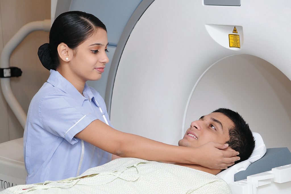
MRI
The advent of MRI has revolutionised the diagnosis and treatment algorithms for sports injuries. The availability of high strength magnets, dedicated coils with improved hardware design and concomitant development of a number of special pulse sequences allows accurate diagnosis of subtle and early bone and soft tissue injuries. Specialised sequences are available for the 3D assessment of articular cartilage. Evaluation is improved by intra-articular instillation (correct word?) of dilute contrast or gadolinium to obtain MR images with exquisite resolution (MR arthrography). At Kokilaben Hospital, these studies are performed on the state-of-the-art 3 Tesla MR equipment with dedicated coils and specialised pulse sequences.
Conclusion
With advancement in radiology, the significance of clinical assessment in deciding when to investigate, what to treat and the implications of abnormal findings is increasing. This is because the incidental anatomical variants or normal developmental findings and asymptomatic degenerative changes that are more frequent with age and high-level sporting activity, are now detected more often with superior resolution offered by modern imaging techniques.
At Kokilaben Hospital, the decision to offer the most beneficial treatment is arrived at after joint discussions between specialist orthopedic surgeons and dedicated musculoskeletal radiologists; this despite the fact that a gamut of sophisticated radiological techniques are at the experts’ disposal.