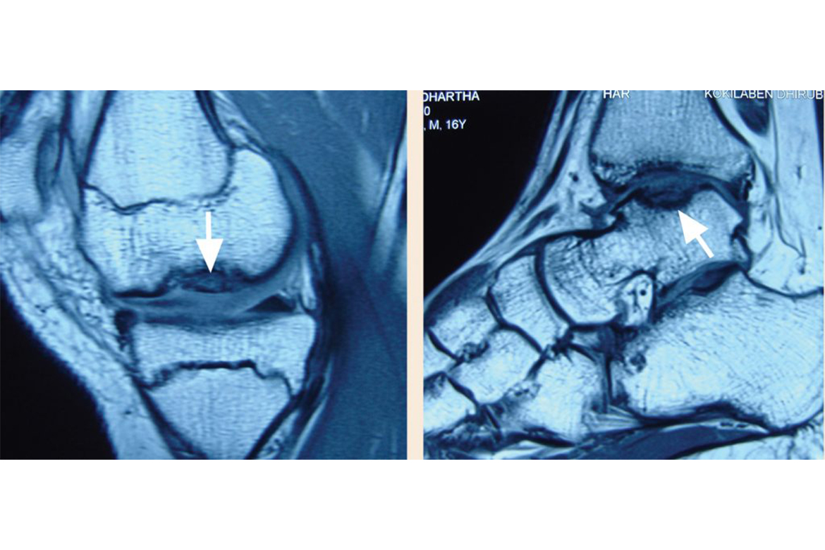
A 16 year-old athlete was referred to the hospital with pain, swelling and episodic locking of the left knee and left ankle which he had twisted during a basketball match two months prior to the consult. He also had a history of pain in both knees, and in the left ankle six months prior to the injury, which he attributed to excessive sports participation.
On clinical evaluation he was found to have an antalgic gait with severe tenderness and effusion in both the left knee and left ankle with mild tenderness in the right knee. Stress provocative tests of the knee demonstrated locking. His radiographs and MRI revealed osteochondritis dissecans of both knees (medial femoral condyle) and left ankle (lateral talar dome).
The left knee and left ankle had advanced stage disease and the separated osteochondral fragment had formed a loose body in the joint. The right knee showed early subclinical disease. A complete haematological work-up to rule out familial multicentric avascular osteonecrosis revealed no abnormality.
Autologous chondrocyte implantation (aci)
ACI is a reconstructive procedure indicated for young patients with focal articular cartilage defects secondary to injury or osteochondritis dissecans. It’s the only procedure that is capable of producing a hyaline-like cartilage repair. It is also more amenable to larger defects than previously described procedures and involves two stages.
- Stage 1:
Arthroscopic evaluation of the focal chondral lesion + chondrocyte biopsy.
The cartilage specimen is sent to the laboratory in a sterile tube with culture medium where the cartilage is enzymatically digested and the chondrocytes isolated. The chondrocytes undergo cell culture for four weeks, which increases the number of cells by a factor of 100.
- Stage 2:
The cultured chondrocytes (usually 48 million cells) are mixed with fibrin glue and this construct is injected directly into the articular cartilage defect. This solidifies within seven minutes of delivery. Unlike older generation ACI, this latest delivery system has the benefits of decreased operating time, smaller incisions, even distribution of chondrocytes, and decreased pain.
The patient is kept non-weight-bearing for six weeks. Patients are typically permitted to return to light sports six months after the surgery.
In a Swedish study, with an average follow-up of ten years, the investigators concluded that a long-lasting, durable cartilage repair is achieved with ACI, and this prevents the early onset of arthritis in patients with focal cartilage defects
What is osteochondritis dissecans (ocd)
Osteochondritis dissecans (OCD) is a joint disorder in which a crack forms in the focal area of the articular cartilage and the underlying subchondral bone causing this osteochondral fragment to become separated or loose from the end of the bone. OCD is caused by blood deprivation in the subchondral bone. This loss of blood flow causes the subchondral bone to die in a process called avascular necrosis. The bone is then reabsorbed by the body, leaving the articular cartilage it supported prone to damage. The result is fragmentation (dissection) of both cartilage and bone, and the free movement of these osteochondral fragments within the joint space, causing pain, locking, and cartilage wear.
OCD occurs most often in young men, particularly after an injury to a joint. The knee is most commonly affected, although it can occur in other joints, including the elbow, shoulder, hip and ankle.
He underwent autologous chondrocyte implantation to reconstruct the osteochondral defects in his left knee and ankle. In the first stage, a knee arthroscopic loose fragment removal and chondral biopsy were performed. Following in-vitro expansion of his chondrocytes, implantation was performed a month later via a mini-open approach in the left knee and via an arthroscopic approach in the left ankle. At present, his chondrocytes are cryopreserved for any possible future implantation that the right knee might require.
It has been over a year since the surgery. The patient has had complete resolution of all symptoms and an excellent return of knee and ankle range and function.
 Back to Site